Paediatric Orthopaedics
Club foot
Clubfoot is a condition in which a child is born with one foot or both feet turned inward (misshaped ). The bottom of the foot may face sideways or even upward.
It is important to start treatment within 1-3 weeks after birth. After corrective bracing, a child may be able to walk, wear shoes, and play sports.
The goal of treatment is to straighten the foot so your child can stand or walk painlessly with the sole of the foot flat on the ground. The foot shape can usually be corrected with stretching, casting, and bracing. In rare cases, surgery is needed.
Treatment with Ponseti Casting (Plaster):
- Gently stretching the foot toward the normal position.
- Holding the foot in place with a full leg cast.
- Removing the cast each week, stretching the foot toward the normal position, and then placing a new cast. This process is continued for 5 to 8 weeks or until the foot is in a normal position.
Your child need to wear a special brace (boots and bar brace) to prevent the deformity from coming back. This brace has both
Flat foot
The condition in which the midfoot sags with lowering of the longitudinal arch of the foot. Flat feet are a common condition, often runs in families and is usually painless. In some cases, flat feet can affect the body’s alignment, which can cause knee and ankle discomfort. Most people with flat feet have no complaints or symptoms of the condition. If the child is having foot pain especially in the heel or arch area that increases with activity, orthopaedic consult is needed. For children who are experiencing pain associated with their flat feet, doctors may recommend a variety of non-surgical treatments to relieve the pain, including arch supports, supportive shoes, stretching exercises, physical therapy and rest. Surgery is not usually needed for flexible flat feet but those with fixed flatfoot in conditions such as Tarsal coalition, surgery may be recommended.
Accessory Navicular
The accessory navicular is an extra bone or piece of cartilage located on the inner side of the foot just above the arch. It is incorporated within the posterior tibial tendon, which attaches in this area and can lead to Accessory Navicular Syndrome. An accessory navicular is congenital (present at birth). Adolescence is a common time for the symptoms to first appear. This is a time when bones are maturing and cartilage is developing into bone. Symptoms include vague pain that usually occurs after a period of activity. Signs include visible bony prominence on the midfoot with swelling and redness. X-rays are usually ordered to confirm the diagnosis. Non-surgical treatment include immobilisation, ice, medications, physical therapy and orthotics. If nonsurgical treatment fails to relieve the symptoms of accessory navicular syndrome, surgery may be appropriate. Surgery may involve removing the accessory bone, reshaping the area and repairing the posterior tibial tendon to improve its function. This extra bone is not needed for normal foot function.
Tarsal coalition
Osteochondritis Dissecans is a condition in which a small segment of bone in a joint begins to separate from its surrounding region due to a lack of blood supply, most often in children and adolescents. The most common joints affected by osteochondritis dissecans are the knee, ankle and elbow. The OCD lesions have a greater chance of separating from the surrounding bone and cartilage, and can even detach and float around inside the joint. In these cases, surgery may be necessary. Symptoms of OCD include pain and swelling of a joint often brought on by sports or physical activity. Advanced cases of OCD may cause joint catching or locking. An x-ray of the affected joint is essential for an initial OCD diagnosis, and to evaluate the size and location of the OCD lesion. Once diagnosis is made, an MRI can help evaluate the extent to which the overlying cartilage is affected. In most cases, OCD lesions in children and young teens will heal on their own, especially when the body still has a great deal of growing to do. Resting and avoiding vigorous sports until symptoms resolve will often relieve pain and swelling. If symptoms do not subside after a reasonable amount of time, your doctor may recommend the use of crutches, or splinting or casting the affected arm, leg or other joint for a short period of time. In general, most children start to feel better over a 2- to 4-month course of rest and nonsurgical treatment. They usually return to all activities as symptoms improve. Surgical treatment is indicated when non-surgical treatment fails; lesion detaches and move around in the joint; lesion is very large (>1cm in diameter). Various surgical techniques include drilling to create pathways for new blood vessel; fixing the lesion with pins or screws; replacing the damaged area with a new piece of cartilage or graft.
Osteochondritis dissecans
Osteochondritis Dissecans is a condition in which a small segment of bone in a joint begins to separate from its surrounding region due to a lack of blood supply, most often in children and adolescents. The most common joints affected by osteochondritis dissecans are the knee, ankle and elbow. The OCD lesions have a greater chance of separating from the surrounding bone and cartilage, and can even detach and float around inside the joint. In these cases, surgery may be necessary. Symptoms of OCD include pain and swelling of a joint often brought on by sports or physical activity. Advanced cases of OCD may cause joint catching or locking. An x-ray of the affected joint is essential for an initial OCD diagnosis, and to evaluate the size and location of the OCD lesion. Once diagnosis is made, an MRI can help evaluate the extent to which the overlying cartilage is affected. In most cases, OCD lesions in children and young teens will heal on their own, especially when the body still has a great deal of growing to do. Resting and avoiding vigorous sports until symptoms resolve will often relieve pain and swelling. If symptoms do not subside after a reasonable amount of time, your doctor may recommend the use of crutches, or splinting or casting the affected arm, leg or other joint for a short period of time. In general, most children start to feel better over a 2- to 4-month course of rest and nonsurgical treatment. They usually return to all activities as symptoms improve. Surgical treatment is indicated when non-surgical treatment fails; lesion detaches and move around in the joint; lesion is very large (>1cm in diameter). Various surgical techniques include drilling to create pathways for new blood vessel; fixing the lesion with pins or screws; replacing the damaged area with a new piece of cartilage or graft.
Osgood Schlatter disease
Osgood-Schlatter disease is a condition that causes pain and swelling below the knee joint, where the patellar tendon attaches to the top of the shinbone (tibia), a spot called the tibial tuberosity. There may also be inflammation of the patellar tendon, which stretches over the kneecap. Osgood-Schlatter disease is most commonly found in young athletes who play sports that require a lot of jumping and/or running. Osgood-Schlatter disease is caused by irritation of the bone growth plate as cartilage in the growth plate is irritated by the prolonged stress resulting in the pain and swelling of the region over tibial tuberosity. Activities that put stress on the knee—especially squatting, bending or running uphill (or stadium steps)—cause the tissue around the growth plate to hurt and swell.
Osgood-Schlatter disease usually goes away with time and rest. Sports activities that require running, jumping or other deep knee-bending should be limited until the tenderness and swelling subside. Kneepads can be used by athletes who participate in sports where the knee might make contact with the playing surface or other players. Ice packs after activity are helpful, and ice can be applied two to three times a day, 20 to 30 minutes at a time, if necessary. The appropriate time to return to sports will be based on the athlete’s pain tolerance. Surgery is recommended only if there are bone fragments that did not heal. Surgery is never done on a growing athlete, since the growth plate can be damaged.
Hip dysplasia
DDH is a condition in which the femoral head has an abnormal relationship to the socket (acetabulum) as a result of improper development of the hip ball and socket joint. The severity may vary from mild dysplasia to frank dislocation of head out of the socket. Severe cases of hip dysplasia are usually diagnosed during a routine screening within the first few months of a child’s life. Other times, the problem may only become noticeable as a child grows and becomes more active during adolescent age.
Causes of hip dysplasia can be due to frank breech delivery, ligamentous laxity or extended hip position in post-natal period. Symptoms include child walking with toe touch due to shortening of the limb. If both hips are dysplastic, child walks with waddling gait with lumbar lordosis and limited abduction. X-rays are done to look for increased acetabular index, disrupted shenton’s line and decreased central edge angle to confirm the diagnosis. In 1 to 6 months of age, conservative treatment is advised in the form of pavlik harness. If reduced on weekly USG, it is continued for 6 weeks followed by abduction orthosis. In 6 to 18 months old child, traction and closed reduction with hip Spica is given for 3 months. If reduction fails, open reduction is recommended. In 18 to 24 months of age, trial of closed reduction or primary open reduction is advised combined with acetabular osteotomy surgery like Salter’s osteotomy. In older child, femoral shortening is used along with above procedures to put less pressure on femoral head after reduction. Hip dysplasia is a treatable condition. However, if left untreated, it can cause irreversible damage of the hip joint and end up in premature arthritis. Therefore, early detection and timely intervention are both important to reduce a child’s risk of pain and disability in adulthood.
Perthes disease
Perthes disease is a rare childhood condition of hip. It occurs when the blood supply to the head of the femur (thighbone) is temporarily disrupted. Without an adequate blood supply, the bone cells die. Perthes is really a complex process of stages that can last several years. As the condition progresses, the weakened bone of the head of the femur gradually begins to collapse.
It occurs in children aged 4 to 10 years, five times more common in boys with 10 -15% involvement of both hips. This is a chronic disease divided into 4 stages which are initial necrosis, fragmentation, reossification and healed stage. Symptoms include child with hip pain, decreased mobility of the hip joint with limping gait.
The goal of treatment is to relieve painful symptoms, protect the shape of the femoral head, and restore normal hip movement. If left untreated, the femoral head can deform and not fit well within the acetabulum (hip socket), which can lead to further hip problems in adulthood, such as early onset of arthritis.
Treatment plan is based on the age of the child and extent of disease involvement. For very young children (2 to 6 years of age) with few changes in the femoral head on x-rays, conservative management is advised in the form of observation, anti-inflammatory medications, physiotherapy and life-style modifications. For older children with more than 50% involvement of the femoral head, surgery is recommended in the form of osteotomy +/- femoral shortening in which bone is cut and repositioned to keep the femoral head snugly in the acetabulum. After the procedure, the child is usually placed in a cast for several weeks to protect the alignment.
SCFE
This condition most often affects the hip in teenagers between the ages of 12 and 16. The condition refers to the slip of the growth center of the hip (the capital femoral epiphysis) on the top of the femur bone. The changes occurring in the growing skeleton during puberty play into the chances that a child will develop SCFE. The cartilage epiphyseal plate is weaker than the surrounding bone. Children who are overweight are more prone to developing SCFE. 20 to 40 percent of the time the condition affects both hips.
Most teenagers with SCFE develop pain in the hip, and they begin to walk with a limp. On examination of the hip, the motion of the hip is abnormal and restricted. X-rays are usually necessary to make the diagnosis in SCFE. Based on X-ray findings of the lateral head-to-shaft angle, the slip can be graded.
The condition can be treated and the complications avoided or reduced if recognized early. Surgery is usually necessary to stabilize the hip and prevent the situation from getting worse. If untreated this can lead to serious problems in the hip joint later in life.
Congenital Muscular Torticollis
The term torticollis means ''twisted neck.'' CMT occurs when there is reduced length and increased tone of the muscle called sternocleidomastoid (SCM) on one side. There are a number of suggested causes, including ischaemia, trauma during childbirth, intrauterine malposition. Child presents with lateral bending of neck towards the affected side with slight rotation of the chin to the opposite side. Diagnosis of CMT can usually be made based on the clinical examination. Ultrasonography (USG) is the most frequently used form of imaging, especially for neonate. It is useful for assessing neck masses, pseudo-tumour and post treatment evaluation. Conservative management include physiotherapy and manual stretching. Surgical management is done in case of no improvement with manual stretching; more than 15 degrees deficit in passive rotation and lateral bending; tight muscular band /tumour in SCM. Surgical options include unipolar/ bipolar SCM muscle lengthening, ‘z’ lengthening.
Bow knees-Genu varum
Genu varum is an angular deformity of the knee in which the child appear ‘’bowlegged’’. Varus is physiological in children aged 6 to 12 years and considered abnormal after 2 years. Pathological causes of genu varum include blount’s disease, nutritional rickets, post traumatic, congenital deformities, fibrous dysplasia. In late adulthood it can progress and lead to osteoarthritis.
Plain radiographs and scannogram are done to see the extent and location of the deformity. Metabolic profile is done to rule out primary underlying cause.
Children with physiological varus are managed by educating and reassuring the parents.
In older children with pathological varus progressively increasing with age, operative treatment is done in the form of tibial osteotomy, femoral osteotomy or guided growth.
Knock knees-Genu valgum
Genu Valgum is a condition in which knees are deviated towards midline of the body and both knees touch each other when straightened.
Physiological genu valgum is present in children upto 4 years. Unilateral genu valgum can be seen in physeal injury, proximal metaphyseal fractures, benign tumour. Both sides genu valgum can be caused by renal osteodystrophy or skeletal dysplasia.
Diagnosis is arrived by examining the patient. X-rays and scannogram are done to calculate the extent and location of the deformity. Non operative treatment is advised in physiological genu valgum in children < 6 years, or treatment of the underlying primary cause. Operative treatment can be done in the form of hemiepiphysiodesis, bone stapling and corrective osteotomy in older children not responding to conservative treatment.
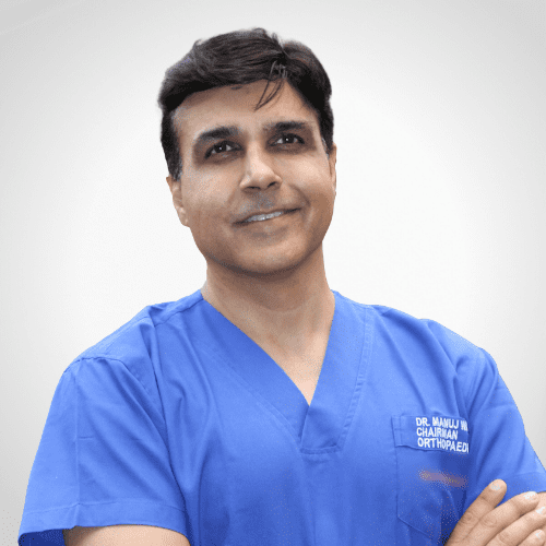
Dr. Manuj Wadhwa
Chairman & Executive Director- Elite Institutes of Orthopedics & Joint Replacement
- Ojas Hospitals, Panchkula
- Ivy Hospitals, Punjab
Awards Wining Doctor
- 2 Times World Book of Records
- 7 Times Limca Book of Records
Contact Us
Let’s Get In Touch
Sector 22, Panchkula
09:00 am - 04:00 pm
(By Prior Appointments)

Sector 71, Mohali
09:00 am - 04:00 pm
(By Prior Appointments)
Email us
Give us a Call
Book An Appointment
We Are Featured In