Arthroscopy and Sports Medicine
ACL reconstruction
A ligament is a strong, cord-like band of tissue that connects one bone to another bone. The anterior cruciate ligament (ACL) connects one of the lower leg bones (tibia) to the upper leg bone (femur).
A completely torn ACL cannot heal on its own. In athletes and other people of any age who wish to continue doing physically demanding activity, ACL reconstruction surgery is needed. ACL reconstruction is a surgery to replace a torn ACL.
It is performed through key-holes (Arthroscopy) via small incisions using a combination of fibre optics and small instruments. In ACL reconstruction surgery, the torn ligament is replaced with a tissue graft, which will mimic the natural ACL. A portion of patient’s own tendon (patellar, hamstring, or quadriceps) is used to make a new ACL. A tunnel is drilled into both the tibia and femur. The graft is threaded across the knee and secured in place by using screws, buttons, staples or sutures. This restores the stability to the knee joint.
Revision ACL reconstruction
A revision ACL reconstruction is a second surgery needed to replace the deficient ACL graft. ACL revision surgery is a more challenging operation than a primary reconstruction. The main reasons a patient might need a revision ACL reconstruction include:
- A re-tear of the ACL
- Problems arising from the previous surgery
- Failure of the reconstructed ligament to heal properly
Revision ACL reconstruction surgery is recommended when there is recurrent instability feeling of the knee, even after focussed rehabilitation and physiotherapy. Some patients may require a two stage revision surgery, in which Bone grafting is done during the first surgery to fill up the previous surgery tunnels in the bones. New ACL ligament is reconstructed using one of the options: the patellar tendon, quadriceps tendon, hamstring tendon, peroneus longus tendon or tendon from the opposite side knee.
Meniscus surgery
Meniscus is operated arthroscopically using small incisions (key-hole surgery) using an Arthroscope, which is a thin tube with light and video camera at its end. The camera projects the video images from inside the knee joint onto a monitor.
Two types of Meniscus Surgery:
- Meniscus Repair – The Torn meniscus portions are stitched together using special instruments and meniscus is allowed to heal.
- Partial Meniscectomy – The damaged meniscal tissue is trimmed and removed leaving the healthy meniscus in place.
Recovery after the Meniscus Surgery
- The recovery time depends on the type of surgery performed.
- Full recovery from Meniscus repair takes between six weeks to twelve weeks.
- After Partial Meniscectomy, the recovery is much quicker.
PCL Reconstruction
- The PCL is a band of tissue that connects the femur to the tibia and stabilises the knee joint from backward translation.
- Isolated PCL injuries of mild grade (grade 1 and 2) can be managed without surgery by Physical therapy and brace.
Significant laxity (grade 3 laxity) & posterior sag or associated injury to other structures like the posterolateral corner (PLC) warrants Surgery – PCL reconstruction. In this Surgery, torn PCL ligament is replaced using a piece of tendon or ligament (autograft). The surgery is performed through key hole incisions (Arthroscopy).
Multi-ligament Reconstruction
Multi-ligament injury of the knee means two or more major ligaments in and around the knee is torn. These injuries always require surgery in the form of arthroscopic and open ligament reconstruction.
The reconstruction requires autografts (patient’s own) harvested from the same or opposite knee or allografts (tissue bank). Generally a waiting period of 10-14 days is required for the soft tissue swelling to subside before surgery. Surgery can be done in single stage or in two stages, depending upon the severity of the injury.
Cartilage restoration Surgeries
An articular cartilage injury is damage to the cartilage that lines the surface of joints (articular cartilage). The cartilage is smooth, white tissue that covers the ends of bones where they meet at a joint. This cartilage allows smooth movement of the joint. It also acts as a thin cushion between the bones of the joint. Injuries to joint cartilage can cause pain and decrease range of movement.
Articular cartilage damage can occur from a recent (acute) injury or from wear and tear that takes place over time. The knee joint is the most common area for this type of injury.
Initial treatment involves rest and analgesics for the relief of pain with supervised physiotherapy. Since the cartilage injuries have very poor healing capacity treatment and prognosis depends on the size of the defect.
For smaller articular cartilage defects which are asymptomatic, surgery may not be required.
Smaller Defects :
Defects smaller than 2 cm have the best prognosis. Treatment of such defects include arthroscopic surgery involving drilling and causing microfractures(creating small holes). This procedure stimulates healing response by marrow cells that produce cartilage at the area of the defect almost similar to the native cartilage.
Larger defects:
For slightly larger defects, it may be necessary to transplant cartilage from other areas of the knee called mosaicplasty/OATS- Osteochondral autograft transfer.
Larger defects:
For very large defects, it is necessary to harvest the patients cartilage cells in the 1st stage operation which are then cultured followed by implantation of these cells about 3 weeks later. This procedure is called autologous chondrocyte implantation(ACI).
Patella Instability Surgery
Patellar instability surgery is a procedure to restrain the kneecap (patella) in its groove (patella femoral groove).
This surgery may involve a combination of procedures:
- Reconstruction of the MPFL ligament using autograft. This is done arthroscopically using key-hole incisions.
- Release of tight tissues on the outer side of your knee.
- Moving the area where your patellar tendon attaches to your shin bone (tibial tubercle transfer).
High Tibial Osteotomy
The knee is divided into two halves, or compartments. The medial compartment is the inside half of the knee and the lateral compartment is the outside half of the knee. Cartilage is the smooth, slippery covering of the bone ends. It allows the bones of the knee joint to slide over each other without rubbing.
The Wear and tear of the articular cartilage (osteoarthritis) sometimes affects one half of the knee far more than the other. In such cases, surgery to realign the lower leg shifts the pressure to the healthier half of the knee. This procedure intends to increase the life span of the joint and prolong the time before a knee replacement surgery becomes necessary.
The procedure involves a cut through the upper part of the tibial bone (Osteotomy). After the bone is cut, the two sides of the tibia are separated to form a wedge-shaped opening. This opening is then filled with bone graft and the bone is stabilised using special plates. This procedure changes the overall axis of the lower leg and shifts the load onto the healthier side within the knee joint. The ideal patient for this osteotomy is a young, active person, who has arthritis limited to one side of the knee joint.
Arthroscopic Loose Body Removal
The procedure refers to the removal of bits of bone, cartilage or other tissue that have broken free and are floating within the joint.
Joint is inspected through key hole incisions using an Arthroscope camera and specialized arthroscopic tools are used to retrieve the loose bodies.
Arthroscopic Rotator Cuff Repair
The rotator cuff is a large tendon comprised of four muscles which combine to form a "cuff" over the upper end of the arm, the head of the humerus. The four muscle are supraspinatus, infraspinatus, subscapularis and teres minor. The rotator cuff helps to lift and rotate the arm and to stabilize the ball of the shoulder within the joint.
A rotator cuff tear may result from an acute injury such as a fall or may be caused by chronic wear and tear with degeneration of the tendon. Surgery is recommended if you have persistent pain or weakness in your shoulder that does not improve with nonsurgical treatment.
Surgery to repair a torn rotator cuff involves re-attaching the tendon to the head of humerus (upper arm bone) using specialized suture anchors. Advances in surgical techniques for rotator cuff repair include less invasive procedures. Surgery is done through small incisions and using Arthroscope and small specialized instruments. Arthroscopic rotator cuff repair causes minimal trauma to the tissues that surround the shoulder and the rotator cuff. Because of this, patients have smaller scars and less damage to nearby structures than open surgery.
Shoulder Instability Surgery
Shoulder Instability results from the tear in the rim of cartilage (labrum) around the edge of the shoulder socket (glenoid). In some cases, bone loss happens at the ball (humeral head) and/or the socket (glenoid)
Recurrent shoulder dislocation almost always needs to be treated with surgery.
Types of surgery for shoulder Instability:
Arthroscopic Bankart repair – This refers to stitching of the torn labrum tissues back to the glenoid socket via key hole procedure. This is recommended in the cases with minimal or no bone loss.
Remplissage procedure – This refers to the filling of the bone defect in the ball (humeral head)
Latarjet procedure – This is recommended for the recurrent shoulder instability with significant bone loss. The procedure involves transferring a small piece of bone called the coracoid process along with its muscle attachments to the socket of the shoulder joint. This compensates for the bone loss and also provide a soft tissue support.
Arthroscopic Shoulder decompression
Arthroscopic decompression is a procedure used to treat shoulder pain due to shoulder impingement syndrome, when this condition has not been relieved with medicines and physiotherapy. In this procedure, the inflamed bursa or bone spur in the shoulder is removed, or shaved away through key-hole incisions using specialized instruments.
This creates more space for the rotator cuff tendons and allows smooth gliding of the rotator cuff under the acromion bone, and enables pain free overhead movements of the shoulder.
AC Joint Reconstruction
Surgery is recommended when the AC joint disruption results in persisting pain or severe deformity.
In this procedure, the AC joint is “reduced” into its normal anatomic position. This can be held with a variety of methods, some requiring metal implants and others require special suture tapes with buttons. The ligaments connecting the collar bone (clavicle) and the shoulder blade (acromion) are reconstructed using the tendon graft harvested from elsewhere in the body.
Biceps Tenodesis
Biceps tenodesis treats biceps tendon tears caused by injury or overuse. The procedure also treats SLAP tears (superior labrum tears of the socket) extending into the biceps tendon. Biceps tendon is located at the top of the biceps muscle. It’s connected to the upper portion of the labrum (rim of cartilage that lines the shoulder socket) In the biceps tenodesis procedure, the torn biceps tendon is released from your labrum and it is relocated to your upper arm bone (humerus) using a special tenodesis anchoring devices through mini-open technique and Arthroscopy.
Frozen shoulder surgery
Frozen shoulder, also called adhesive capsulitis, causes pain and stiffness in the shoulder. Over time, the shoulder becomes very hard to move. If your symptoms are not relieved by therapy and other conservative methods, surgical treatment might be recommended. Manipulation under anaesthesia - During this procedure, your shoulder is made to move after putting you to sleep. This causes the capsule and scar tissue to stretch or tear. This releases the tightening and increases range of motion. Shoulder arthroscopy. In this procedure, tight portions of the joint capsule are released through arthroscopy. This is done using pencil-sized instruments inserted through small incisions around your shoulder. Tight capsular tissues are released using special radio-frequency probe. In many cases, manipulation and arthroscopy are used in combination to obtain maximum results. Most patients have good outcomes with these procedures.
Achilles Tendon Repair
Achilles tendon repair is a surgery to repair an Achilles tendon that has been torn (ruptured). The Achilles tendon is a cord-like band that connects the muscles of your lower leg (calf) to your heel. Achilles tendon facilitates walking by helping to raise the heel off the ground. Mid-substance tears:
Various surgical techniques are available to repair the rupture. In acute tear end to end suturing of the torn ends are done through mini-open technique. Chronic and retracted tears often require lengthening or augmentation by other tendons
Insertional tears: The procedure involves the excision of the bony bump(Haglund’s bump) and repair of the tendon back to the bone using special suture anchors.
Ankle Ligament Reconstruction
An ankle ligament injury is a stretch or tear in a ligament in the ankle. Ligaments are tissues that connect bones to each other.
This condition is often caused by rolling or twisting the ankle. Most of these are mild disruptions in the ligament (sprains) and heal fully within 6 weeks of injury with proper treatment. If the ligaments are completely torn, patients may develop persistent instability (give way) feeling of the ankle joint. Chronic ankle instability is a condition that makes the ankle weak, painful, and more likely to give way.
Ankle ligament reconstruction is a procedure to fix a torn or stretched ligament in the ankle. The most common operation is called a Brostrom¬ Gould lateral ligament reconstruction, where the ligaments are repaired through an incision on the outer aspect of the ankle. The procedure also involves a key hole surgery to assess the joint cartilage. The ligaments are repaired back to the bone using specialised suture anchors and supplemented with suture tapes.
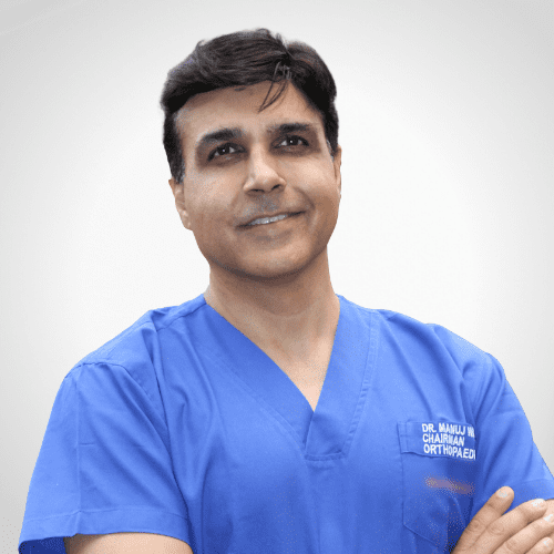
Dr. Manuj Wadhwa
Chairman & Executive Director- Elite Institutes of Orthopedics & Joint Replacement
- Ojas Hospitals, Panchkula
- Ivy Hospitals, Punjab
Awards Wining Doctor
- 2 Times World Book of Records
- 7 Times Limca Book of Records
Contact Us
Let’s Get In Touch
Sector 22, Panchkula
09:00 am - 04:00 pm
(By Prior Appointments)

Sector 71, Mohali
09:00 am - 04:00 pm
(By Prior Appointments)
Email us
Give us a Call
Book An Appointment
We Are Featured In